Friday, August 31, 2012

Reading a CBC, nRBC's / Nucleated RBC's


Nucleated red blood cells are immature RBC's that haven't lost their nucleus yet.  These cells are called metarubricytes and look like this:


These immature red blood cells are usually pushed out early in response to anemia, so they indicate regenerative anemia.  They are not usually found in normal animals.

Reading a CBC, WBC / NCC / White Blood Cell Count

NCC stands for "Nucleated Cell Count" which works as "White Blood Cell Count" for mammals, but not for birds or reptiles which have nucleated red blood cells.

This is usually a machine-generated number although the total white blood cell count can be done manually with a hemocytometer as well.

White blood cells include neutrophils, lymphocytes, monocytes, eosinophils, and basophils but collectively they are responsible for what we generally think of as immunity.  They range from highly specialized cells that go after particular antigens to generalized eaters of our body's "garbage" or stuff like absorbable suture that the body identifies as foreign and, therefore, in need of disposal, or even cells responsible for allergic reactions.

Increases in white blood cells or "leukocytosis" indicates:
-Excitement: this is common in cats.  If they become overly excited during the blood draw all of the cells that are normally loosely attached to the endothelial lining of the blood vessels detach and end up in circulation where they can be drawn out and counted.  This means that the increase isn't "real" because there aren't really more cells being made, it's just that the ones that aren't usually counted because they stick to the blood vessel wall were counted even though in 30 minutes or so they'll go back to where they belong.  If this is the case you'll see normal or slightly increased lymphocyte numbers.
-Inflammation or other causes of neutrophilia:  because neutrophils are the most common leukocyte (at least in dogs and cats), when the numbers of neutrophils increase dramatically you can also end up with a leukocytosis as a result.

-Decreases in white blood cells or "leukopenia" indicates:
- Viral diseases: such as feline distemper (aka "panleukopenia"), canine parvovirus, etc
-Infection: bacterial infection, septicemia, toxemia, endotoxins
-Drug-related: such as with the use of sulfonamides

Reading a CBC, Basophils

Basophils may be counted by machines or by a technician doing a manual differential or "manual diff".  This involves taking a drop of blood and making a smear on a slide.  It is allowed to dry, then stained with Diff Quick stain.  Once the stain has dried the slide is viewed under a microscope and evaluated for red and white blood cell morphology as well as a count of 100 white blood cells to determine what percent of cells are of each WBC type including monocytes.

Basophils are rare.  If you see one while looking at a slide, consider yourself lucky.  If you hear someone else say they found one, ask to see it.  I have yet to see a basophil myself.

Below is a photo of a normal basophil:


Basophils are associated with eosinophils and mast cells, but their use on a CBC is limited due to how rare they are.  The causes of their increase are unknown, but usually associated with eosinophilia, or an increase in eosinophils as well.

Reading a CBC, Eosinophils

Eosinophils may be counted by machines or by a technician doing a manual differential or "manual diff".  This involves taking a drop of blood and making a smear on a slide.  It is allowed to dry, then stained with Diff Quick stain.  Once the stain has dried the slide is viewed under a microscope and evaluated for red and white blood cell morphology as well as a count of 100 white blood cells to determine what percent of cells are of each WBC type including eosinophils.

The number of eosinophilss may be expressed as a percent or as an absolute number which is expressed as some number per microliter.  If you have a choice it is best to work with the absolute number since, with abnormal WBC counts, the number expressed as a percent can be misleading.

Below is a photo of a normal eosinophil although the size of the red granules differ with species, especially the can in which they are more pale and rod-shaped:


Eosinophils are responsible for allergic responses and also play a part in systemic parasite infestations.

Increases in eosinophils or "eosinophilia" occurs as a result of:-Worms: systemic parasites such as heartworm or hookworms.
-Weases: allergic reactions or hypersensitivity reactions including flea bite dermatitis.
-Weird diseases:  feline hypereosinophilic syndromes and some poorly characterized gastroenteritis cases.

Decreases in eosinophils or "eosinopenia" occurs as a result of:-Stress:  due to increased natural or administered steroids.

Reading a CBC, Monocytes

Monocytes may be counted by machines or by a technician doing a manual differential or "manual diff".  This involves taking a drop of blood and making a smear on a slide.  It is allowed to dry, then stained with Diff Quick stain.  Once the stain has dried the slide is viewed under a microscope and evaluated for red and white blood cell morphology as well as a count of 100 white blood cells to determine what percent of cells are of each WBC type including monocytes.

The number of monocytes may be expressed as a percent or as an absolute number which is expressed as some number per microliter.  If you have a choice it is best to work with the absolute number since, with abnormal WBC counts, the number expressed as a percent can be misleading.

Below is a photo of a normal monocyte:


Monocytes are the cleanup crew for the body.  They migrate out of the blood vessels as needed and then become tissue macrophages, which means they eat dead tissue and anything else that doesn't belong in the body.

Increased monocytes or "monocytosis" is caused by:
 -Inflammation: either acute or chronic
-Stress: due to natural or administered steroids, especially in dogs.

Decreased monocytes or "monocytopenia" is caused by:
-Pancytopenia: usually an incidental finding when all white blood cell counts are low which is called "pancytopenia".

Reading a CBC, Lymphocytes

Lymphocytes may be counted by machines or by a technician doing a manual differential or "manual diff".  This involves taking a drop of blood and making a smear on a slide.  It is allowed to dry, then stained with Diff Quick stain.  Once the stain has dried the slide is viewed under a microscope and evaluated for red and white blood cell morphology as well as a count of 100 white blood cells to determine what percent of cells are of each WBC type including lymphocytes, which are the most common white blood cells in cattle.

The number of lymphocytes may be expressed as a percent or as an absolute number which is expressed as some number per microliter.  If you have a choice it is best to work with the absolute number since, with abnormal WBC counts, the number expressed as a percent can be misleading.

Below is a photo of 2 normal lymphocytes:


Lymphocytes include the cells responsible for both humoral and cell-mediated immunity.  Sometimes they may appear with blue cytoplasm, these are reactive forms which are likely B cells.

Increased lymphocytes or "lymphocytosis" is caused by:
-Excitement: this is common in cats.  If they become overly excited during the blood draw all of the cells that are normally loosely attached to the endothelial lining of the blood vessels detach and end up in circulation where they can be drawn out and counted.  This means that the increase isn't "real" because there aren't really more cells being made, it's just that the ones that aren't usually counted because they stick to the blood vessel wall were counted even though in 30 minutes or so they'll go back to where they belong.  If this is the case you'll see increased numbers of neutrophils as well (neutrophilia).
-Leukemia


Decreased lymphocytes or "lymphocytopenia" is caused by:

-Stress: an increase in steroids which can also be due to the administration of steroids
-Acute viral infection: usually accompanied by beutropenia or a decrease in neutrophils as well.

Reading a CBC, Segs / Neutrophils

Neutrophils may be counted by machines or by a technician doing a manual differential or "manual diff".  This involves taking a drop of blood and making a smear on a slide.  It is allowed to dry, then stained with Diff Quick stain.  Once the stain has dried the slide is viewed under a microscope and evaluated for red and white blood cell morphology as well as a count of 100 white blood cells to determine what percent of cells are of each WBC type including neutrophils, which are the most common white blood cells in dogs and cats.

The number of neutrophils may be expressed as a percent or as an absolute number which is expressed as some number per microliter.  If you have a choice it is best to work with the absolute number since, with abnormal WBC counts, the number expressed as a percent can be misleading.

Below is a photo of a normal, mature, segmented neutrophil:


Neutrophils are the body's first line of defense against infection and also play a part in inflammation.

Increased neutrophils is called "neutrophilia" and may indicate:
-Inflammation:  if this is the case it is often accompanied by a left shift.
-Stress: caused by an increase of steroids including those used as medication and usually indicated by a decrease in lymphocytes aka "lymphocytopenia".
-Excitement: this is common in cats.  If they become overly excited during the blood draw all of the cells that are normally loosely attached to the endothelial lining of the blood vessels detach and end up in circulation where they can be drawn out and counted.  This means that the increase isn't "real" because there aren't really more cells being made, it's just that the ones that aren't usually counted because they stick to the blood vessel wall were counted even though in 30 minutes or so they'll go back to where they belong.  If this is the case you'll see normal or slightly increased lymphocyte numbers.

Decreased neutrophils is called "neutropenia" and may indicate:
-Inflammation: usually accompanied by a left shift.
-Infection: especially viruses such as parvovirus.  In this case other blood cell numbers will also go down in the order of their circulating lifespan.
-Bone marrow suppression: due to illness or drugs such as estrogen, chemotherapy, ehrlichiosis

Reading a CBC, Bands

Machines cannot reliably distinguish between band neutrophils and mature neutrophils yet (although they are very close), so this value is usually determined by a technician doing a manual differential or "manual diff".  This involves taking a drop of blood and making a smear on a slide.  It is allowed to dry, then stained with Diff Quick stain.  Once the stain has dried the slide is viewed under a microscope and evaluated for red and white blood cell morphology as well as a count of 100 white blood cells to determine what percent of cells are of each WBC type including bands.

Bands are immature neutrophils.  In the photo below, the white blood cell on the left is a band neutrophil while the one on the right is a "Seg" or normal, mature, segmented neutrophil:


Immature "band" neutrophils are released from the bone marrow when the body is running low on circulating, mature, neutrophils and decides that immature cells are better than none at all.  Neutrophils are the body's first line of defense against infection, but they are also involved in inflammation.  So, whichever the cause, band neutrophils indicate that the body's normal defenses aren't able to keep up.

When bands are present it's called a "left shift".  The next question to ask yourself is whether there are more bands or more mature neutrophils present.  If there are more mature neutrophils than bands it is a better prognosis and it's called a "regenerative left shift".  This means that bands are being produced, but the body is still putting up a good fight.  If there are more bands than segs, it's called a "degenerative left shift".  This is a bad prognosis because it indicates the body is not keeping up -it is losing the battle.

The presence of bands indicates:

-Inflammation:  this is the most common cause.
-Bone marrow disorders such as leukemia may cause early release of neutrophils.
-Bone marrow injury

The absence of bands indicates:
-Normal, yay!

Reading a CBC, Reticulocytes

Reticulocytes are young red blood cells.  If viewed on a slide with Diff Quick stain they'd be called polychromatophils because they'd appear larger than the RBC's and slightly bluish in color.  When viewed on a slide using New Methylene Blue stain they appear as large RBC's with one or more dark purplish dots inside.  These are the remnants of the nucleus which is not quite gone because of the cells immaturity.  The image below is of reticulocytes stained with New Methylene Blue:


If reticulocytes are present it may indicate:
-regenerative anemia

If reticulocytes are not present it is:
-normal!  Yay!

Reading a CBC, MCHC / Mean Corpuscular Hemoglobin Concentration

MCHC or Mean Corpuscular Hemoglobin Concentration is the average amount of hemoglobin in the red blood cells.  It is usually a machine-determined value, but it can be estimated like this:
MCHC = (Hb / PCV) X 100

Increased MCHC:
-Machine error: there is no such thing as hyperchromic RBC's

Decreased MCHC:-Regenerative anemia: immature cells have less hemoglobin than mature cells, so a low MCHC can indicate regenerative anemia.  MCV, reticulocytes, and nRBC's should also be looked at as well to confirm that the cause of the low MCHC is regenerative anemia.

Reading a CBC, MCV or Mean Corpuscular Volume

MCV stands for Mean Corpuscular Volume.  In plain English that's the average size of the red blood cells.  It is usually determined by machine, but can be calculated manually like this: 
MCV = (PCV / RBC count) X 10.

Increased MCV or "macrocytosis" may be due to:
-Regenerative anemia:  Immature RBC's are larger than mature RBC's, so a high MCV is one indicator of regenerative anemia although reticulocytes and nRBC's should also be looked at.
-Clumped platelets:  Clumps of platelets may be counted as RBC's by the CBC machine.  If this is suspected a slide should be made from the same blood sample that was used to run the CBC in order to check for clumped platelets.

Decreased MCV or "microcytosis" may be due to:

-Extreme spherocytosis: Usually spherocytosis does not result in a decreased MCV, but in extreme cases it can.

Reading a CBC, Hgb / Hb / Hemoglobin

Hemoglobin is often abbreviated Hgb or Hb, either way it is the measurement of the oxygen-carrying component of the red blood cells.  This number should be pretty close to being equal to the PCV divided by 3.  I remember this because it rhymes: "Hb equals PCV over 3".  This value is determined by the CBC machine.

Increased hemoglobin indicates:
-Machine error: there is no such thing as a real increase of hemoglobin.

Decreased hemoglobin indicates:
-Regenerative anemia: young RBC's don't have as much hemoglobin as mature cells.  See MCV, reticulocytes, and NRBC's to confirm.

Reading a CBC: RBC / Red Blood Cell Count

The Red Blood Cell Count is given by a CBC machine and tells you the number of RBC's in a microliter of blood.  This number should be compared to the PCV since they both are giving you an idea of the number of red blood cells in the blood. 

Increased RBC count may be caused by:
-Platelet clumping: platelet clumps are often read by machines as being RBC's.  If there are a lot of clumps it could throw your RBC count off by quite a bit.  Compare to your PCV and check your MCV to determine if this is the case.
-Dehydration: check the albumin, if it is also high dehydration is likely.


Decreased RBC count may be caused by:
A decreased RBC count indicates anemia which has 3 main causes:
-Lack of RBC production: from bone marrow issues, nutrition problems, or kidney problems since the kidneys stimulate production via erythropoietin.
-Increased RBC destruction: due to intracellular parasites, immune disorders (IMHA), etc
-Loss: from external or internal bleeding.

Reading a CBC, PCV / HCT


PCV is the Packed Cell Volume which is also known as the hematocrit or HCT.  Generally this is determined by taking a microhematocrit tube, filling it with blood collected in a tube, plugging one end with clay, and then spinning it at very high speeds in a centrifuge.  After spinning, the blood components will separate in the tube with the red blood cells on the bottom, the white blood cells on top of that, and the plasma at the very top.  The hematocrit tube is then placed on a reading card as shown below with the top of the clay lined up with the bottom line and the top of the plasma lined up with the top line.  Finally, you find the number that matches up with the top of the red blood cells in the tube.  So, in the picture below, if the red lines indicate changes of 10% on the table, the PCV of that hematocrit tube would be about 50%. 


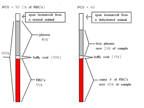
It is important to keep in mind that what this test really measures is the percent of the blood that is made up of red blood cells.  The PCV is expected to go down in cases of anemia, but go up in cases of dehydration since there is less water (plasma) but the same number of RBC's.  The drawing below may make the change with dehydration more clear.  Click on it if you need to see a larger version of the image.

PCV changes w/ dehydration
Decreased PCV:
A lower than normal PCV indicates anemia which has 3 main causes:
-Lack of RBC production: from bone marrow issues, nutrition problems, or kidney problems since the kidneys stimulate production via erythropoietin.
-Increased RBC destruction: due to intracellular parasites, immune disorders (IMHA), etc
-Loss: from external or internal bleeding.

Increased PCV:
-Dehydration: this is most common, but it is important to look at the albumin results of the Chemistry test and compare it to the PCV.  If both are high the cause is likely dehydration.